
Amazing collection of microscopic pictures from inside of human body provided by Wellcome Images, UK shows various parts of human body up close and personal. Most of the pictures have been color enhanced to help important part stand out and get distinguished from its surrounding. This also adds to more 3D feel of the pictures that have depth thanks to color adjustments. Human body is truly amazing.
Vast maority of pictures has been photographed using SEM – scanning electron microscope which emits high energy beams that scan surfaces and create microscopic images of it as it “scans” across the cells. SEM can send millions of beams across the target area. Those slid along the surface of atoms present and send information that is then processed into a high resolution, 3D image. SEM can create high-res images of any part of human body and magnify it 250,000 times, which means it can create a high resolution image even of atoms as small as 1nm (nano meter).
The history of SEM images reaches back to 1935 when Max Knoll captured first microscopic picture of silicone steel. The SEM technology has since evolved and improved to a level that now it can create a nice, clean 3D image of even the smallest part of human body. Following microscopic pictures will take you on a journey through human body, showing detailed images of neurons in human brain, through plaque on teeth, down to surface of a healthy lung and a lung suffering from cancer, all the way to human egg, sperm and an embryo fertilized by sperm.
It’s a fantastic world of human body up close and personal. Great images with lots to offer. After you have seen these microscopic pictures, you may never look at human body the same again. All pictures were released by Wellcome Images under the Creative Commons License. Authors include Yorgos Nikas, Dr. David Becker, Professor Alan Boyde, Anne Weston, David Gregory & Debbie Marshall, Freya Mowat, Annie Cavanagh and Liz Hirst. Enjoy the human body from the inside in a series of 15 amazing, microscopic pictures.
-

- Human Body Microscopic Picture – Egg Coated with the Zona Pellicuda and Two Coronal Cells Attached to it
-
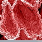
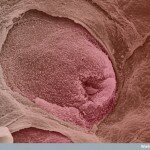
- Human Body Microscopic Picture – Villi of Small Intestine
-
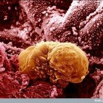
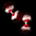
- Human Body Microscopic Picture – Sperm Trying to Fertilize a Human Egg
-
- Human Body Microscopic Picture – Retinal Blood Vessels Branching from the Optic Nerve
-

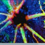
- Human Body Microscopic Picture – Purkinje Neurons in the Brain
-
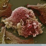
- Human Body Microscopic Picture – Lung Cancer Cell
-
- Human Body Microscopic Picture – 6 Days Old Embryo inside Woman’s Womb
-

- Human Body Microscopic Picture – Stereocilia Hair inside the Ear
-
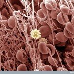
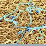
- Human Body Microscopic Picture – Blood Clot with White Blood Cell
-
- Human Body Microscopic Picture – Alveoli Cavities in the Lung Tissue
-
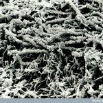
- Human Body Microscopic Picture – Tooth Plaque
-
- Human Body Microscopic Picture – Taste Bud on the Tongue
-

- Human Body Microscopic Picture – Red Blood Cells
-
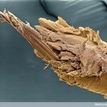
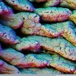
- Human Body Microscopic Picture – Damaged End of Human Hair
-
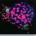
- Human Body Microscopic Picture – Human Embryo Surrounded by Sperm

You can post this "Human Body from the Inside - Amazing Microscopic Pictures" image that's above on your blog/forum using following codes:
Website Code
Forum Code


Most People Reading This Article Found It Searching For:
- Inside Human Body
- cells in the human body
- Picture Inside Human Body
- Michael Crichton
- microscopic photo of tounge
- human body pictures
- inside human body pictures
- amazing human body
- pictures of the human body
- microscopic images of human hair
ooh! what a fantastic image. Is the colors original or …created.
THE ACHIEVEMENT AND GIFT OF SCIENCE
FOR SCIENCE!
Amazing pictures, GOOD is Grate. It is way to find
ABSOLUTELY ASTOUNDING PHOTOS!
WHO CAN DOUBT THERE IS A GOD?
these are truly amazing brings more “insight” to what I am learning about in school